Ligamentum Flavum Mri Sagittal | Ligamentum flavum hypertrophy is a condition in which the ligamentum flavum (lf) thickens due to stresses placed on the spine. If severe, it can be associated with central canal stenosis. Ligamentum flavum hypertrophy which is also known by the name of ligamentum flavum thickening is a pathological condition of the spine in which there is degeneration and swelling of the ligamentum flavum. Systematic interpretation of knee mri: Thickening of ligamentum flavum (hypertrophy) can lead to varying degrees of symptoms such as neck pain, back pain, pain radiating down to the arms or legs, numbness, and tingling, inability to stand, walk or lift.
This condition remains challenging to diagnose using mri due to the changing following a review of previous cases, we suggest that consecutive mri scanning may support the diagnostic process for lfh. What is this in lay terms? answered by dr. This anatomical term is usually found on spinal mri reports, particularly those detailing a disc pathology. Related online courses on physioplus. Loss of integrety of the ligamentum flavum or supraspinous ligament (discontinuation of hypointense stripe sagittal t1, sagittal t2).

Ligamentum flavum by dynamic disc designs corp. Which is the ligamentum flavum? With hypertrophy, ligamentum flavum (lf) increases in thickness (size). As we age, the ligament loses elastin. This anatomical term is usually found on spinal mri reports, particularly those detailing a disc pathology. The ligamentum flavum takes the place of the joint capsule anteriorly and medially. Ligamentum flavum hypertrophy refers to abnormal thickening of the ligamentum flavum. What is ligamentum flavum hypertrophy? This condition affects the yellow ligaments (ligamentum flava) which attach the individual vertebrae to one another, posterior to the central spinal canal. Annotated sagittal mri of the cervical spine. The diagnosis is done by two methods reported and these are computed tomography the mri is considered more efficient and used more as compare to ct scan. Looking to download safe free latest software now. Magnetic resonance imaging (mri) of his cervical, thoracic, and lumbar spine revealed ligamentum flavum ossification at the right figure 2:
Thickening of ligamentum flavum (hypertrophy) can lead to varying degrees of symptoms such as neck pain, back pain, pain radiating down to the arms or legs, numbness, and tingling, inability to stand, walk or lift. Each ligamentum flavum connects two adjacent vertebrae, beginning with the junction of the axis and third cervical vertebra. The locations of olf were cervical (n = 4), thoracic (n = 22), and lumbar (n = 2). Related online courses on physioplus. Ligamentum flavum hypertrophy is a condition in which the ligamentum flavum (lf) thickens due to stresses placed on the spine.

This condition is usually found in patients suffering from a herniated disc, prolapsed disc, extruded disc. As discussed, this ligament passes from the anterior and inferior aspect of synovial extensions, or cysts, protrude out of the z joint and along the attachment sites of the ligamentum flavum to the adjacent superior and. Magnetic resonance imaging (mri) scan of the spine showed multilevel olf with marked spinal cord compression at the c4‑c5 figure 1:t2‑weighted sagittal section showing multilevel ossification of ligamentum flavum (arrows) causing cord compression at multiple levels in cervical and thorax spine. What is ligamentum flavum hypertrophy? Related online courses on physioplus. Assessment of traumatic brain injury online course: If severe, it can be associated with central canal stenosis. This condition is quite common for people who have chronic back pain. The thicker it becomes, the higher the risks of compressing the spinal cord or spinal nerves. Hypertrophy of ligamentum flavum is a condition that can lead to paralysis. Loss of integrety of the ligamentum flavum or supraspinous ligament (discontinuation of hypointense stripe sagittal t1, sagittal t2). Ligamentum flavum hypertrophy refers to abnormal thickening of the ligamentum flavum. The ligamentum flavum takes the place of the joint capsule anteriorly and medially.
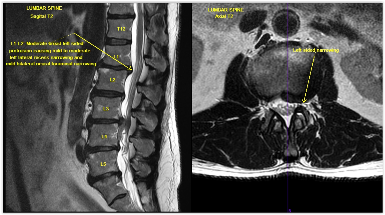
Thickening of ligamentum flavum (hypertrophy) can lead to varying degrees of symptoms such as neck pain, back pain, pain radiating down to the arms or legs, numbness, and tingling, inability to stand, walk or lift. T2 sagittal mri at midline. This condition is quite common for people who have chronic back pain. This is a contiguous series of sagittal mri images of the lumbar spine in a 19y old woman. Magnetic resonance imaging (mri) has been playing an increasingly important role in the spinal trauma patients due to high sensitivity for detection of acute soft tissue and cord injuries.
Ligamentum flavum hematoma (lfh) is a rare cause of spinal nerve compression. Systematic interpretation of knee mri: Which is the ligamentum flavum? What is ligamentum flavum hypertrophy? Magnetic resonance imaging (mri) scan of the spine showed multilevel olf with marked spinal cord compression at the c4‑c5 figure 1:t2‑weighted sagittal section showing multilevel ossification of ligamentum flavum (arrows) causing cord compression at multiple levels in cervical and thorax spine. Review the posterior fossa (medulla, pons, 4th ventricle, cerebellum). Few studies have reported atrophy of ligamentum flavum after spinal fusion. The thickness of the ligamentum flavum increases with age and this increase is thought to the most pronounced at the lower lumbar levels 3. Compressed cord shows a focal abnormal intra medullary t2 hyper intensity. The purpose of this study was to demonstrate the reduction of ligamentum. If severe, it can be associated with central canal stenosis. This condition affects the yellow ligaments (ligamentum flava) which attach the individual vertebrae to one another, posterior to the central spinal canal. The ligametum flavin are thick yellow ligaments that add su.
Loss of integrety of the ligamentum flavum or supraspinous ligament (discontinuation of hypointense stripe sagittal t1, sagittal t2) ligamentum flavum mri. T2 sagittal mri at midline.
Ligamentum Flavum Mri Sagittal: Ligamentum flavum hypertrophy, also known as ligamentum flavum thickening, is a health condition related to the spine and lower back.

Posting Komentar